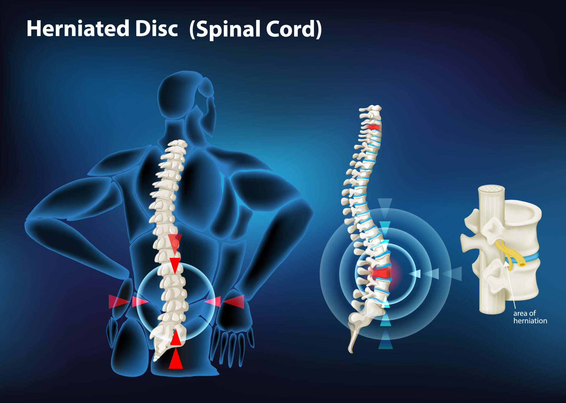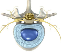
The spine comprises of a column of vertebral bones intervening with intervertebral discs. (See diagram on the right) These discs play multiple roles including – shock absorption, allowing spine flexibility and movement, maintain disc height and contributing to spinal stability. In this blog, we discuss about the anatomy of the intervertebral disc, annular tears (a common cause of low back pain in young patients) as well as the treatment of this very painful condition.
Anatomy of the Intervertebral Disc

Annular Tear
Symptoms of an Annular Tear
Diagnosis of an Annular Tear
An annular tear causing low back pain is diagnosed via a combination of clinical evaluation, imaging studies and diagnostic injections. Clinical evaluation is based on history as well as features in your physical examination. The best imaging modality to appreciate an annular tear is a Magnetic Resonance Imaging.
Not all annular tears are symptomatic. Therefore, the radiological findings of an annular tear does not mean that it is necessarily the cause of the pain. If there is significant doubt regarding the cause of the pain, your doctor may recommend that you undergo a test known as provocation discography. This study involves injection of the affected disc to see if the pain can be replicated.
Treatment of Low Back Pain from an Annular Tear
The treatment of Annular tears follows the principles of the management of low back pain. While the care of the patient is highly individualized, most patients receive conservative treatment at the start. If the pain is well controlled with conservative treatment, no further action is necessary. Examples of conservative treatments include:
- Medications
- Physical therapy
- Rest and activity
- modification
- Preventing of re-injury (eg. limit weight lifting, twisting activites)
- Weight management
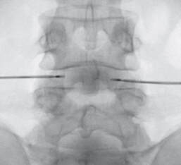
In some patients, the pain persists for weeks to months after conservative treatment. In these patients, more invasive treatments may be warranted. A commonly performed treatment is Annuloplasty. In this procedure, needles are placed in the intervertebral discs and heat is applied across the discs. This procedure helps to denervate the disc and interrupt the barrage of pain signals entering the brain during activity. It can also help to seal up the annular tear in a significant proportion of patients who undergo this procedure.


